Wednesday, September 22, 2010

Anatomy of an electric ray

I love my job. Sometimes I just get to participate in (or watch) cool stuff. As was the case a week ago, when I learned that we were going to be dissecting an electric ray.

This electric ray had been brought in by a fisherman, who didn't want to eat it or let it go to waste. So we decided to hold a public dissection, so that we could learn more about it. We also preserved some of the pieces for our collection, to be used for later research or teaching.


Electric rays are slow-moving rays that live off of the pacific cost, in cool waters. They don't have a stinger, but do have a specialized organ that produces electricity, which they use to capture food and defend themselves.

In this picture you can see the two-chambered heart, center, at the right most edge of the cut. Below that (center) is the stomach. To either side of the stomach are two very large livers. Like all sharks and rays, the electric ray lacks a swim bladder and depends on the oil reserves in the liver to help maintain buoyancy. The large green ball near the upper liver lobe is the gall bladder.


This is a nice close-up of the electric producing organ. Basically, it is little more than coin shaped muscle stacks. Since muscles produce electricity, the arrangement of muscle tissue in this configuration helps optimize the amount of electricity produced in the area. By having an organ on either side of the body, the electric ray can stun prey trapped between the two sides of their fins.

Tuesday, August 31, 2010

Slime star


This is Pteraster tesselatus, commonly known as a slime star. As its name implies, when disturbed this star can produce copious amounts of slime, which may protect it from predators [1]. It eats sponges and the like and lives in the colder waters off the west coast of the US.

It also has an interesting feature common to many members of its family. It has a 'brood pouch'. The surface that you see is actually a soft covering which covers the true surface of the sea star. In other members of the family, females will release their eggs from their gonopores (like all other sea stars), but retain them under that covering until they develop into juvenile sea stars and crawl out. In this particular species, they do not brood their young, and instead send them shooting out of the osculum, an opening in the covering. The osculum is generally used for exchanging water from the water vascular system to the outside, and can be seen opening and closing even in individuals who are not spawning.

Thursday, August 19, 2010

My little squids

Well, the class is wrapping up, which is why I have not been able to post much. We did two projects which required a lot of sleepless nights. Luckily, I was able to finish my paper last night and actually went to bed before 1 am. I did a little study to see if I could reduce the number of embryos in squid egg capsules to see if it affected development. I was quite pleased with it, because they actually survived (not a sure thing when you are breaking open egg capsules).


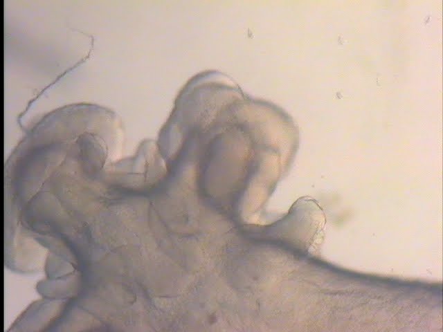
So here is a pic from one of my squids...


If you look closely at the top of its mantel disk, you can see its two little bumps that will become the fins. The arms /tentacles are forming, and the eyes are becoming more defined and developed. And my favorite part... on its arms are little suckers!


They've still got a bit to go, but since tomorrow is the last day, I've returned the rest of the mass back where it was collected.

Friday, August 13, 2010

I have eyes!


Or at least eye primordia. This is a picture of one of the squid embryos I am working with for my second project. If you look closely at the lumps on either side of the embryo you'll see clear ovals, that's were the eyes will develop. I am very excited to see how they will look tomorrow!

It's been a bit busy, as the paper for my first project was due at the start of the week, at the same time as the proposal for my second (and a presentation on the first during the middle of the week). Hopefully, I'll get a good head start on the second paper tonight... so I can take the time to go whale watching this weekend. I can't believe the program is ending so soon.

Monday, August 2, 2010

Microplankton and Megaplankton!


Another member of the plankton. This is a larval ctenophore. You can make out its ctene rows along the side of the body and the massive mouth right in the center. When I see them, they normally have a copepod stuffed inside.

On the macro side, we found this large fried egg jelly (Phacellophora camtschatica) floating around our dock the other day. The bell can get up to 2 ft (60 cm) in diameter and the tentacles 20 ft (6 m) long. They eat other jellyfish.