Showing posts with label development. Show all posts

Sunday, December 4, 2011

Mysterys and metamorphosis

I am being more and more drawn to the mysterious bug world that seems to be invading my life. Now I can experience the thrill of invertebrates on land! When I noticed this strange bug the other day I was completely taken aback. I could not figure out even what kind of bug this could possibly be. I suspected that it might be something larval, as I saw a few shed exoskeletons which might have been the same thing. But still, what was it? Not a caterpillar that was for sure. It had six legs, so it must be an insect... not a spider. But no wings? Hum...

To top it off, I noticed yet another strange bug near by. It looked like it might have been a beetle, but there was no hardened wing coverings and the abdomen was just to darn long. And what was with those crazy 'feet' at the end of the abdomen?

Puzzled I took some photos and went on my way, thinking I would never know for sure. I tried some searches for my mystery bug number one, and thought it might be some kind of mealy bug, but I was not totally convinced.

So a few days pass, and I go out to my plants where mystery bug number two is hanging out and low and behold I see a new bug, this one fairly recognizable:

Well, well a ladybug hanging out where mystery bug number two is... Maybe mystery bug number two is a larval form of the lady bug. They are the same color, and you can see the spots starting to form. So I do a little searching and find out that this lady bug is Harmonia axyridis, an asian ladybug. Lucky for me, I also get to solve the identity of mystery bug number two AND number one.

As I suspected, mystery bug number two is a pupal form; of the asian ladybug. Number one is the larva of that same individual. What I took for 'feet' was actually the discarded molt of the larva at the end of the pupa. Metamorphosis is so cool... but I can imagine that it drove early entomologists up the wall trying to identify things, much like it drives us plankton-lovers up a wall!

Friday, July 8, 2011

Abalone development


I recently got to be a part of an abalone spawning team, where we induced abalones to release their gametes. It was very fun to see the fertilized eggs develop, and interesting for me because it was the first time that I had actually seen spiral cleavage in action! It was so different looking than radial cleavage...

I did not manage to get pictures of all of the stages, as I had to sleep, so expect some gaps. But I hope you enjoy this abalone development slide show that I put together of the stages I did see.

Monday, June 27, 2011

Grunion Development: Day 9


Not much seems to have changed from day 8 to day 9. The grunion is still the same size, and has not gained any new pigmentation. The yolk has decreased a bit, but at this point no major developmental; changes are visible. However, on day 9 they are much easier to hatch!


You can still see the remnants of the yolk in the young fish's stomach.

Sunday, June 26, 2011

Grunion Development: Day 8


The eyes are fully pigment, and the body as well. The body has grown to such a length that it wraps around the inside of the egg a little over 2 times. The oil droplets have been completely depleted, and there is a little yolk left. What you can't see is that the embryo is very active, with eye and body twitches being quite common.

Friday, June 24, 2011

Grunion Development: Day 6


The embryo has grown, with the tail wrapping all the way around the egg and back around the head. The eyes have a lot of pigment and the body is starting to get a little pigment. The oil droplets are almost all used up and the yolk has a lot of blood vessels connecting it to the embryo.

Wednesday, June 22, 2011

Grunion Development: Day 4


The embryos are noticeably larger, and structures are becoming readily apparent. You can see the nerve cord running down the center of the embryo. gill arches forming behind the eye, and the eye itself has an indented pupil.

What you can't tell from the picture is that the heart is also nociable and pumping, and there are musculature visible along the length of the tail.

Tuesday, June 21, 2011

Grunion Development: Day 3


Now you can clearly see many features of the developing embryo. It has also grown large enough to wrap around the back of the egg, with the tail just peeking out on the left hand side. The oil droplets are being used up and condensed into one large drop right in the center.

Monday, June 20, 2011

Grunion Development: Day 2


Now it's approximately 37 hours after fertilization. You can still see the oil droplets off to the left, but the embryo has under gone some big changes. Instead of being a cap of cells it now has a definite form, with the head and developing eyes in the center, and the rest of the body trailing off to the top right.

Sunday, June 19, 2011

Grunion Development: Day 1


I thought I'd take some pictures of grunion eggs as they develop, just because I could. Here is day 1, approximately 13 hours after fertilization. You can see the dark orange oil droplets in the center, and a pale yellow cap of cells off to the left. That is the developing embryo.

Wednesday, January 19, 2011

Story of a black jelly


Last summer we had a rare event. Tens of black sea nettles (Chrysaora achlyos) washed ashore on our beach. These guys are pretty rare, so we jumped at the chance to get some gonadal tissue to start a new culture of jellies. We collected the adults from the beach, and I extracted the gonadal tissue from the insides of the bell. (This was a rather painful process which involved me getting stung for three days.)

I used bits of the tissue to sex the animals; females had eggs, and males had packets of sperm. After I figured out who was what, I put a little bit of male gonads and female gonads together in a petri dish and mixed them up, to beak open the male's sperm packets. (It felt a bit like making red scrambled eggs.)

Male sperm packets


Female tissue with eggs


After 3 days, planulae were spotted swimming in the petri dishes.


After 5 days, the planulae settled to the bottom to become polyps. These are newly settled with only 2 fully formed tentacles.


Four months later, I am very happy to say that my polyps have begun to strobilate. I have some beautiful ephyrea, that will become (in my opinion) the prettiest-colored jellies ever.

Thursday, August 19, 2010

My little squids

Well, the class is wrapping up, which is why I have not been able to post much. We did two projects which required a lot of sleepless nights. Luckily, I was able to finish my paper last night and actually went to bed before 1 am. I did a little study to see if I could reduce the number of embryos in squid egg capsules to see if it affected development. I was quite pleased with it, because they actually survived (not a sure thing when you are breaking open egg capsules).


So here is a pic from one of my squids...


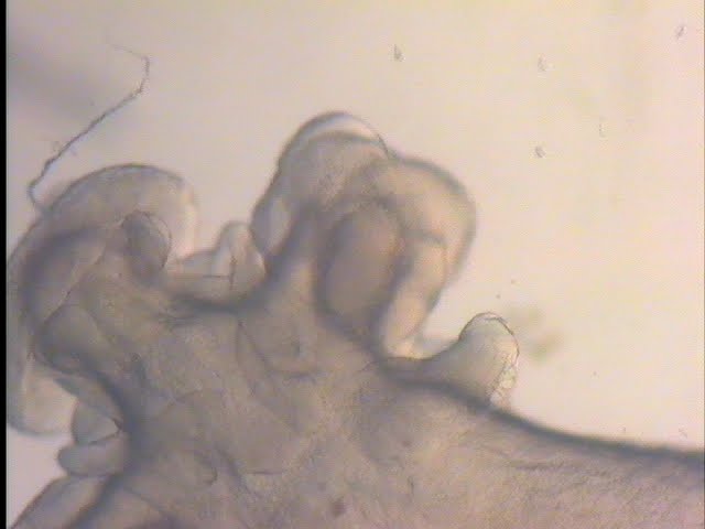
If you look closely at the top of its mantel disk, you can see its two little bumps that will become the fins. The arms /tentacles are forming, and the eyes are becoming more defined and developed. And my favorite part... on its arms are little suckers!


They've still got a bit to go, but since tomorrow is the last day, I've returned the rest of the mass back where it was collected.

Sunday, August 1, 2010

Coolest thing ever?! I think so!


So we're encouraged to have pets in class, and I chose to take care of some Owenia sp. because I thought their development was cool. Today I was changing their water and giving the more food, when I thought I'd check them out under the scope. Some of them were really close to settlement, and even started settling on the slide. Luckily, there are tons of video cameras around and I was able to capture the process.

Here it is, Owenia settlement, speed up, in all its glory.

Saturday, July 24, 2010

Friday Harbor


So, I have just gone up to Friday Harbor Laboratories and have been enjoying my immersion in science after quite a long spell without. I am learning all about larval biology, so have been going on field trips to many different sites, like this mudflat pictured above, to look at egg masses and collect larvae.

One of my favorites is this Owenia sp., which is a type of worm that builds tubes out of small sandy particles. This creature has an interesting development, as the juvenile worm develops around the intestine of the larval body. When it is ready to settle, it drops out of the sac where it was developing and eats its old larval body.

Thursday, March 19, 2009

Life Photo Meme: Quiet


Kingdom: Animalia

Phylum: Chordata

Class: Actinopterygii

Order: Atheriniformes

Family: Atherinopsidae

These are grunion (Leuresthes tenuis). Found off the coast of California, these fish breed from March to July. Approximately three days after the full and new moons, these fish throw themselves on the sandy beaches of southern CA to lay their eggs. Any noise causes them to quickly flop back into the ocean.

Females bury themselves upright in the sand, while males lay on top of the sand next to the females. After the eggs are laid in the sand and fertilized, both sexes return to the ocean. The female can lay up to 3,000 eggs on a single night, and will return to the beach each time the tide is right.

Eggs take about 9 days to develop, but won't hatch until the waves from the high tides break them open, about 12 to 14 days later. Here is a picture of the developing eggs at about 6 days of age. You can already see the well-developed eyes.


If you were to look at these eggs under a microscope you can also see small red blood cells flowing through the blood vessels covering the yolk. You can also see the heart beating ( in this video, it looks like the heart is above the eyes). The tail and its associated muscles are also well developed and serve to move the small fish around in its egg.

Thursday, November 6, 2008

Life photo meme: Mystery Sacoglossan


Kingdom: Animalia

Phylum: Mollusca

Class: Gastropoda

Order: Sacoglossa

Family: Hermaeidae

I recently went on a collecting trip up the coast, and there were hundreds of these sea slugs wandering around on the mud flats we were at. They are really small (my hand is the background in the picture), and the trip leader could not identify them. I took a quick picture and they took a egg mass back with them, to try and ID these slugs. I asked a friend who works with sea slugs and she tentatively identified them as an Aplysiopsis species. Interestingly, she had gone on a trip farther north, and seen hundreds of these as well. So they seemed to be blooming all along the central/northern part of the coast.

Alas, we won't truly know what species it is unless we look at its radula and penis, two of the main features used to distinguish different sea slugs. But this little adventure has allowed me to get some fun footage of mystery slug veliger larvae! This larval type is characteristic of gastropods, and if you look at them you will notice that as larvae, they all have shells. The shell is lost in the adult sea slug.

Without further ado...

Sunday, June 22, 2008

Two layers or three?

Most creatures in the animal kingdom have three cell layers; endoderm, ectoderm, and mesoderm. Mesoderm is particularly important in increasing complexity, as many of our internal organs are derived from this layer. So when did this layer arise?

To look at this question many researchers have turned to cnidarians (jellyfish & anemones), which only have two layers, to examine the developmental and genetic clues. To make things confusing, some cnidarians posses a third layer, called a entocodon, where some muscle cells reside. Also, all ctenophores (comb jellies) also posses muscle cells. Muscle cells are generally thought to have arisen from mesoderm. So was the ancestor to cnidarians, ctenophores, and bilaterians (everybody else) triploblastic (having three layers), and the cnidarians and ctenophores just lost that layer? Or was the ancestor diploblastic (having two layers) and muscle cells arose separately in all groups?

Looking at some of the genes that are commonly associated with mesoderm, Martendale et al. (2004) found that a majority of these genes are present in his model anemone, and generally tend to be expressed in the endoderm. This means that the tools for creating mesoderm was present in cnidarians, and that most likely, mesoderm arose from endoderm at some later date. However, finding the genes in the endoderm does not completely rule out the possiblity that cnidarians had mesoderm, but that it was lost at a later date.

Burton (2008) reviewed the two possibilities, diploblastic or secondarily diploblastic through mesoderm loss, and makes several excellent points based on numerous papers. First, the third tissue in some cnidarians (entocodon), is not the same genetically or developmentally as mesoderm. The entocodon arises from the ectoderm at a much later developmental time (after gastrulation) than mesoderm. Plus, genes associated with mesoderm are not always expressed in the entocodon, they are more likely to be expressed in the endoderm. So the entocodon is most likely a new cell layer and not a modified mesoderm layer.

Muscle cells found in ctenophores and cnidarians are not the same as those found in bilaterians or even to each other, however the genes are similar. Therefore, it is likely that the genes for muscles were found in the ancestors to ctenophores, cnidarians, and bilaterians and each group slightly modified those genes to get their present shape. In triploblasts, these genes along with others became associated with the mesoderm cell layer, when those cells migrated from the endoderm. Interestingly, in cnidarians the genes associated with mesoderm in bilaterians appear to be used in body patterning. So essentially, cnidarians have two sets of body patterning genes, one of was free to develop into mesoderm and subsequently, internal organs. Even cooler, (I think) ctenophore lack one set of body patterning genes, the Hox genes. Did they lose it? Or did they never have it and we are more closely allied with cnidarians? Do sponges and placazoans have Hox genes? I guess it's time for more reading!

Burton, P.M. 2008. Insights from diploblasts; the evolution of mesoderm and muscle. Journal of Experimental Zoology Part B-Molecular and Developmental Evolution. 310B:5-14

Martindale, M.Q., K. Pang, and J.K. Finnerty. 2004. Investigating the origins of triploblasty: 'mesodermal' gene expression in a diploblastic animal, the sea anemone Nematostella vectensis (phylum, Cnidaria; class, Anthozoa). Development. 131:2463-2474

Saturday, December 8, 2007

Weekly Video: Fertilization envelope formation

Furious bouts of writing followed by furious bouts of procrastination (or is it preceded by?). My newest draft of thesis intro is done (I think this is version seven...), and now I am on to rewriting the methods section...tomorrow. Here is a video of a fertilization envelope forming around a sand dollar egg. Next semester, I might re-film it at a higher magnification.


Saturday, November 3, 2007

Weekly Video: 1st Division

It's my favorite lab to teach, development! Here is a video of the first division of a fertilized sand dollar embryo. It's a little choppy, I was trying to figure out how to speed it up in the new iMovie program and was only partially successful.

Thursday, November 1, 2007

Development: how big is big enough?


This picture is of a fertilized sand dollar egg. You can see the dark pigment dots and the clear circle around the egg, which is the fertilization membrane, and the light smears which are sperm still trying to get at the egg. However, the fertilization membrane prevents any other sperm from entering the egg.


I read an interesting paper about development and sea urchins. A little background: there are three different developmental modes in many different invertebrate species. One is nonfeeding, where the young develop into adults using only the energy (yolk) provided by the mother. Another is feeding where the young spend more time in the larval stage feeding and that’s where they get the energy to transform into the adult. The final mode is very rare and it is called facultative feeding. In this case the young don’t have to feed to transform, but they can to get extra energy. Unlike the nonfeeding larvae who can’t feed even if they wanted to, because they don’t have a complete digestive track.


As you can imagine egg sizes for these different mode vary. Since the nonfeeding modes rely only on what the mother supplies to get to a suitable spot and change into an adult, they tend to be very large. The feeding ones are smaller, because they only have to have enough energy to create a gut, then they can feed themselves till they become an adult. The facultative ones are in between (in general). It is thought that this mode is a very unstable one, and that the species that have it are on their way from feeding to nonfeeding modes or vice versa.


What these researchers tried to do was to take a facultative feeder and force it to become a feeder by reducing the amount of energy available to the young. They did that by taking 2-cell and 4-cell stage and breaking them apart, so that one treatment had ½ the energy and the other treatment had ¼ the energy.


2-cell stage 4-cell stage


They then raised some of each type with food and without food to see if the young with less energy were eating and growing faster than those with less energy who had no food. Of course, they found that young that had not been manipulated were larger that those that had been halved or ‘fourthed’. What was interesting was that none of the size-manipulated young were forced to feed to complete metamorphosis. All young transformed at the same time!
This means that the mothers gave up to 4 times the amount of energy needed to undergo metamorphosis to their young!


For more info see the original paper: Allen, J.D., C. Zakas, R.D. Podolsky. 2006. Effects of egg size reduction and larval feeding on juvenile quality for a species with faciltative-feeding development. Journal of Experimental Marine Biology and Ecology. 331:186.197

Wednesday, October 17, 2007

Smells like Home (repost)


So I just got back from a great meeting, and now that my brain is functional again, I wanted to share an interesting talk that I attended. Basically, for those who have never experienced it, when you got to a meeting you get to here a ton of research, most of which is not published yet or in progress. So several days of hearing really smart people doing really cool stuff! (although it can be a mixed bag…) My brain exploded. But now that the knowledge has begun to settle…
The following is a description of some work being done by several people in Australia.


Okay, some background. Most fish have a stage after thy hatch when they float around in the water, before they get big enough to settle out. This would lead to mixing of fish, as they could potentially end up anywhere. However, when they looked at the genetics of some of these fish found on near by reefs, they found that they were very different and not mixing. This means that the baby fish are finding away to keep themselves in the area, or coming back when they are big enough to swim. But how do they know which reef is the one they hatched from?
These researchers looked at larval fish just before they became big enough to settle and tried to see if the babies could ‘tell’ their home by the smell of the water. What they found was that babies found on one reef preferred the water of that reef to any others. They also found that when they kept the babies in the different water for a time to get them to adjust to the new water, they still preferred their ‘home’ water.


Since most of these fish were brooded as eggs on the reef, the researchers were also curious to see when this smell impression was made. Were they impressed while they were in the egg or at hatching? So they took some anemone fish eggs (clown fish), which were known to home not only to reefs, but also back to particular anemones, and ran some tests. Some eggs were kept in anemone water till just before they hatched, and others were only put in anemone water when they were hatching. What they found was that the fish imprinted on the anemone, smelling water immediately after hatching, but not while they were in the eggs.

What it means is that it is possible that some of the fishes on reefs smell the water after hatching, than use the odor to keep themselves from getting too far from their home. So when it comes time for them to settle, they settle on the same area they were born in!
Below is the original abstract with the names and the affiliations of the researchers working on this. I look forward to reading the paper when it gets published.


MILLER-SIMS, V*; ATEMA , J; GERLACH, G; KINGSFORD, MJ
Boston University Marine Program, Marine Biological Laboratories, James Cook University

Olfactory imprinting in coral reef fish

Most marine organisms have a pelagic larval dispersal phase, leading to the question of how far larvae disperse. Larval behavior and odor preferences may play an important role in larval dispersal and settlement. Apogonid larvae prefer the odor of the reef on which they were caught over other reefs and ocean water. It is possible that this response is due to acclimatization to the odor of water the fish have been recently swimming instead of a long-term preference. We tested apogonid larvae settling on One Tree Island by catching them as they came onto the reef and testing their preference for water for One Tree vs. water from Heron Island in a flume preference test. We then held the fish in either One Tree or Heron water and tested them over a period of nine days. The preference for One Tree water declined in both groups over time; there was no significant difference between the animals held in One Tree or Heron water and both groups maintained a preference for One Tree throughout the testing period. Odor preferences remain stable over time despite exposure to other odors and it is possible they are the result of olfactory imprinting to the home reef odor. Olfactory imprinting has been shown in anemonefishes, but the sensitive period is unknown. Breeding pairs of Amphiprion melanopus were held either with or without an anemone. Eggs and larvae were exposed the anemone from egg laying through hatching (1), from egg laying to just prior to hatching (2), just previous to and 1 hour after hatching (3) or had no anemone exposure (4). At 15 days those larvae in groups 2 and 4 had no preference for the anemone while those in groups 1 and 3 showed a strong significant preference for anemone odor. In this species of reef fish larvae must be exposed to the imprinting odor after hatching in order to learn it.